A complete list of publications from the lab can be found on our google scholar page. Feel free to request PDFs of publications by emailing mkersh at Illinois dot com.
Here you can find a summary of recent publications.
 | Minton DM, Ailiani AR, Focht MDK, Kersh ME, Marolf AJ, Santangelo KS, Salmon AB, Konopka A. The common marmoset as a translational model of age-related osteoarthritis. GeroScience, 1- 21, 2024. | 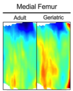 | Three-dimensional reconstructions of subchondral thickness on the medial femur of adult (left) and geriatric (right) marmosets. Blue: low thickness Red: high thickness |
|
| Studying age-related osteoarthritis, which results in both cartilage and bone damage, in humans is difficult due to an unpredictable onset. Working with collaborators at University of Wisconsin-Madison, Michael Focht analyzed bone in a natural animal model of osteoarthritis (OA) finding increased cortical and trabecular thickness in geriatric versus adult marmosets. This now establishes the possibility of using the marmoset as a model – without the need for invasive induction of OA – to help identify treatments to prevent cartilage loss. | ||||
 | Pineda Guzman RA, Naughton N, Majumdar S, Damon B, Kersh ME. Assessment of mechanically induced changes in helical fiber microstructure using diffusion tensor imaging. Annals of Biomedical Engineering, 1-13, 2023. | 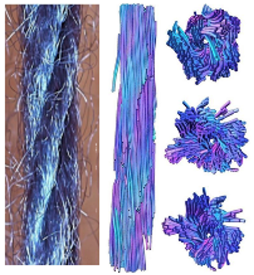 | MRI reconstruction (right images) of the twisting patterns of helical fibers (left photograph). | |
| Noninvasive methods to detect healthy from damaged tissues in vivo are needed. Diffusion Tensor Imaging (DTI) is an imaging technique that can infer tissue microstructure. Working with imaging scientists at the Beckman Institute and the Carle-Illinois Advanced Imaging Center, Roberto Pineda Guzman assessed changes caused by mechanical loading in tissue-mimicking helical fiber constructs using 9.4T DTI data. These results provide evidence of the ability of DTI to detect changes in tissue microstructure that are not apparent at the bulk level, thus confirming its potential as a diagnostic tool for ligament and tendon injury. | ||||
 | Halloran K, Focht MDK, Teague AJ, Peters J, Rice I, Kersh ME. Moving forward: A review of continuous kinetics and kinematics during handcycling propulsion. Journal of Biomechanics, 159, 111779, 2023. | 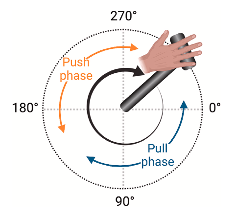 | Illustration of the push and pull phase during handcyling. | |
| As we prepared to begin a new project collaborating with Joseph Peters and Prof. Ian Rice of the Wheelchair Biomechanics Lab, Kellie Halloran, Michael Focht, and CIMED medical student Alex Teague undertook a systematic review of the literature to determine what is known about the continuous kinetics and kinematics during hand cycling. This helped our group to identify gaps in the literature and kickstart our own efforts to understand the biomechanics of hand cycling to evaluate whether it is a viable exercise option for manual wheelchair users. | ||||
| Halloran K, Peters J, Focht MDK, Rice I, Kersh ME. Propulsion kinetics of recumbent handcycling during high and moderate intensity exercise. Journal of Biomechanics, 156, 111672, 2023. Video of handcyling during pilot testing of project. | ||||
| As part of a larger project, Kellie Halloran designed an instrumented handle for a recumbent handcycle which allowed for continuous force measurements. With help from Michael Focht and collaborator Joseph Peters, we characterized three-dimensional continuous applied forces at the handle during high intensity and moderate intensity training. These applied forces can give an early indication of joint loading, and therefore injury risk, at the shoulder. As expected, the forces during HIIT were larger than those in MICT throughout exercise. Throughout the exercises, the timing of peak forces was later in HIIT compared to MICT and could indicate either altered kinematics or muscular fatigue at the end of the exercise protocol. Joseph Peter’s work focused on analyzying the cardiorespiratory response to exercise and participate perception of exercise: Peters J, Halloran K, Focht MDK, Huang K, Kersh ME, Rice I. Cardiorespiratory responses to an acute bout of high intensity interval training and moderate intensity continuous training on a recumbent handcycle in people with spinal cord injury: A within-subject design. Topics in Spinal Cord Injury Rehabilitation, 29 (4),16-26, 2023. Peters J, Halloran K, Teague AJ, Abou L, Erlenbach E, Kersh ME, Rice I. Perceptions of high intensity interval training among people with spinal cord injury: A mixed-methods analysis. Adapted Physical Activity Quarterly, in press, 2023. |
||||
 | Moshage SG, McCoy AM, Kersh ME. Elastic modulus and its relation to apparent mineral density in juvenile equine bones of the lower limb. Journal of Biomechanical Engineering, 145(8), 1-13, 2023. Video showing reconstruction of CT data to evaluate bone properties. See full article here. | |||
| Density–modulus relationships are necessary to develop finite element models of bones that may be used to evaluate local tissue response to different physical activities. It is unknown if juvenile equine trabecular bone may be described by the same density-modulus as adult equine bone, and how the density-modulus relationship varies with anatomical location and loading direction. To answer these questions, Sara Moshage mechanically tested trabecular bone cores from the third metacarpal (MC3) and proximal phalanx (P1) bones of juvenile horses. These samples were from foals that died of natural causes and were acquired with the help of equine surgeon Dr Annette McCoy. We found that density-modulus relationships for juvenile equine trabecular bone were significantly different for each anatomical location (MC3 versus P1) and orientation (longitudinal versus transverse). Moving forward, more accurate models of young bone can now be developed and used to evaluate potential exercise regimens designed to encourage bone adaptation. | ||||