We study the mechanics of bone during postnatal development and aging, and how disease affects structure-mechanical function relationships in bone. This work often incorporates loading on the bone from muscles in order to understand the response of bone to different activities.
| CURRENT WORK | Previous work in bone can be found on our publications page | ||
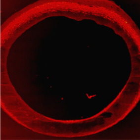
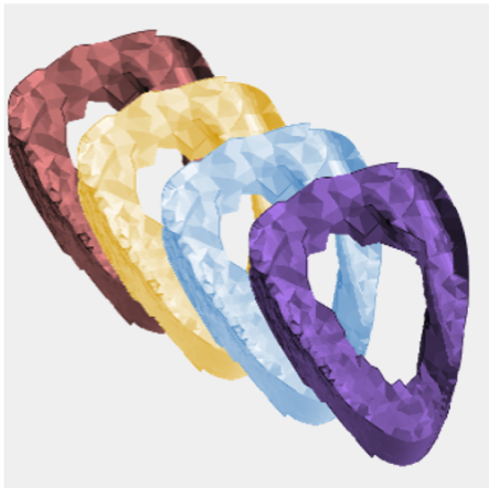
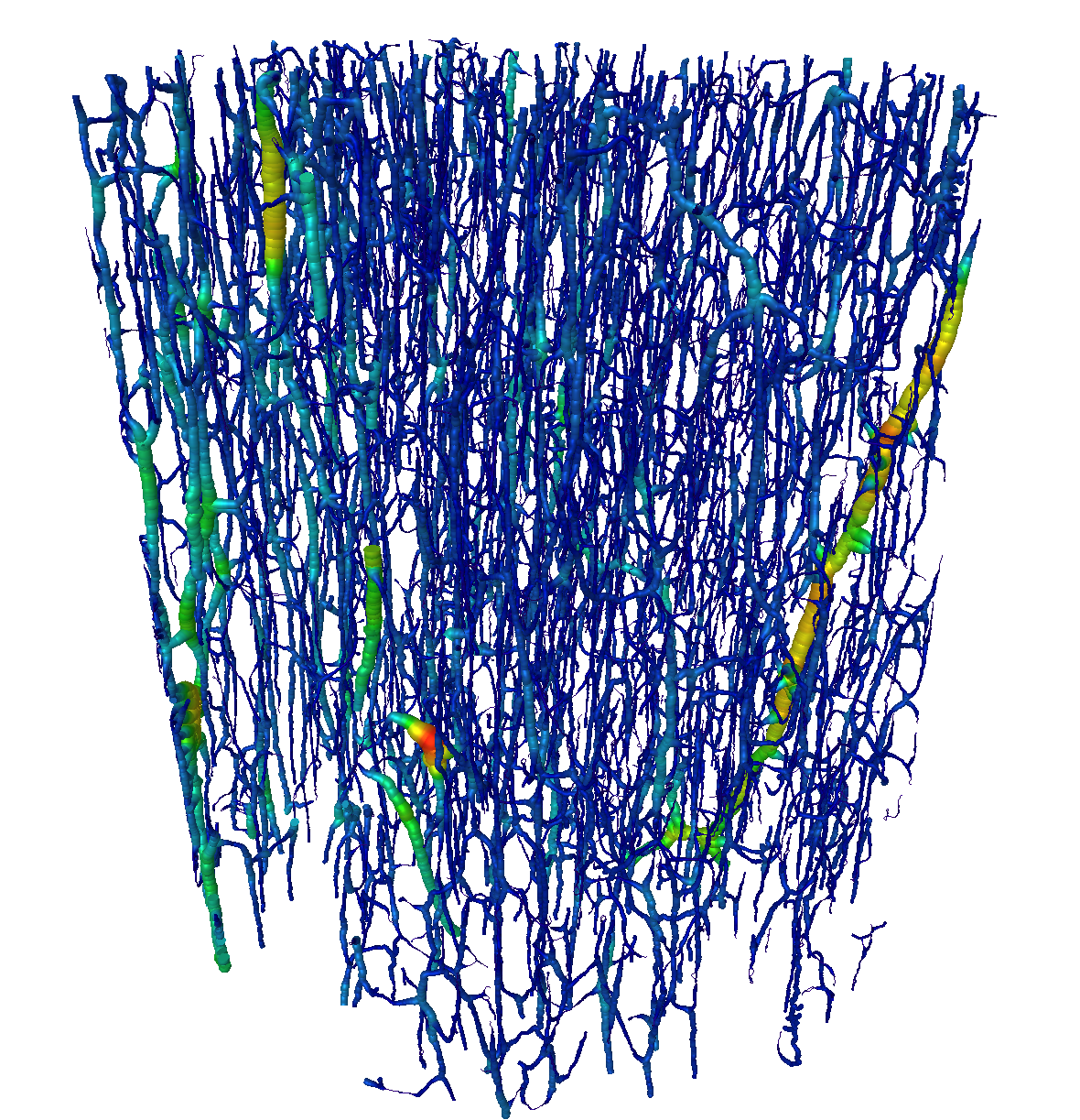 | Microstructure and Strength of Cortical Bone Femoral neck fractures are unfortunately common and lead to high disability and mortality rates. Most fractures are attributed to the onset and accumulation of damage in cortical bone, which is the dense outer layer of bone. Our aim is to understand whether the formation of the pore network of cortical bone (see image) is influenced by the local mechanical environment. We relate cortical bone structure to strength using a combination of ex-vivo, x-ray computed tomography (CT) imaging, mechanical testing, and finite element (FE) modelling. Contributors: Mikayla Hoyle, Mike Rogalski, Liz Livingston, Lydia Bakalova Collaborators: Professor Thomas Andersen (University of Southern Denmark, Cell Biology) Funding: Velux Foundation, UIUC MechSE Department, Burroughs Wellcome Fund | Project lead: Mikayla Hoyle 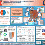 ORS-2024-Poster |
|
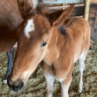 | Myokines and Their Relationship with Bone Remodeling Bone (re)modeling during growth and in response to exercise is a complex process, and a better understanding of the factors that influence remodeling could lead to more effective strategies to prevent fractures. It is well known that the contraction of skeletal muscle during locomotion stimulates bone by exerting forces, but muscle contraction may also provide a biochemical stimulus for bone (re)modeling via the expression of myokines. The overall aim of this research is to elucidate the biological crosstalk between muscle and bone in the context of an early exercise intervention in foals. The research will identify the myokines correlated with bone remodeling as well as the levels of myokines in vivo in horses that correlate with bone structural and compositional properties, muscle forces, and normal growth. Contributors: Samantha Hammack, Sara Moshage Collaborators: Professor Annette McCoy (UIUC, Veterinary Medicine), Professor John Polk (UIUC, Anthropology) Funding: UIUC VetMed Companion Animal Research Foundation, UIUC VetMed Hatch Grant, Morris Animal Foundation, Campus Research Board, Grayson Jockey Club |
||
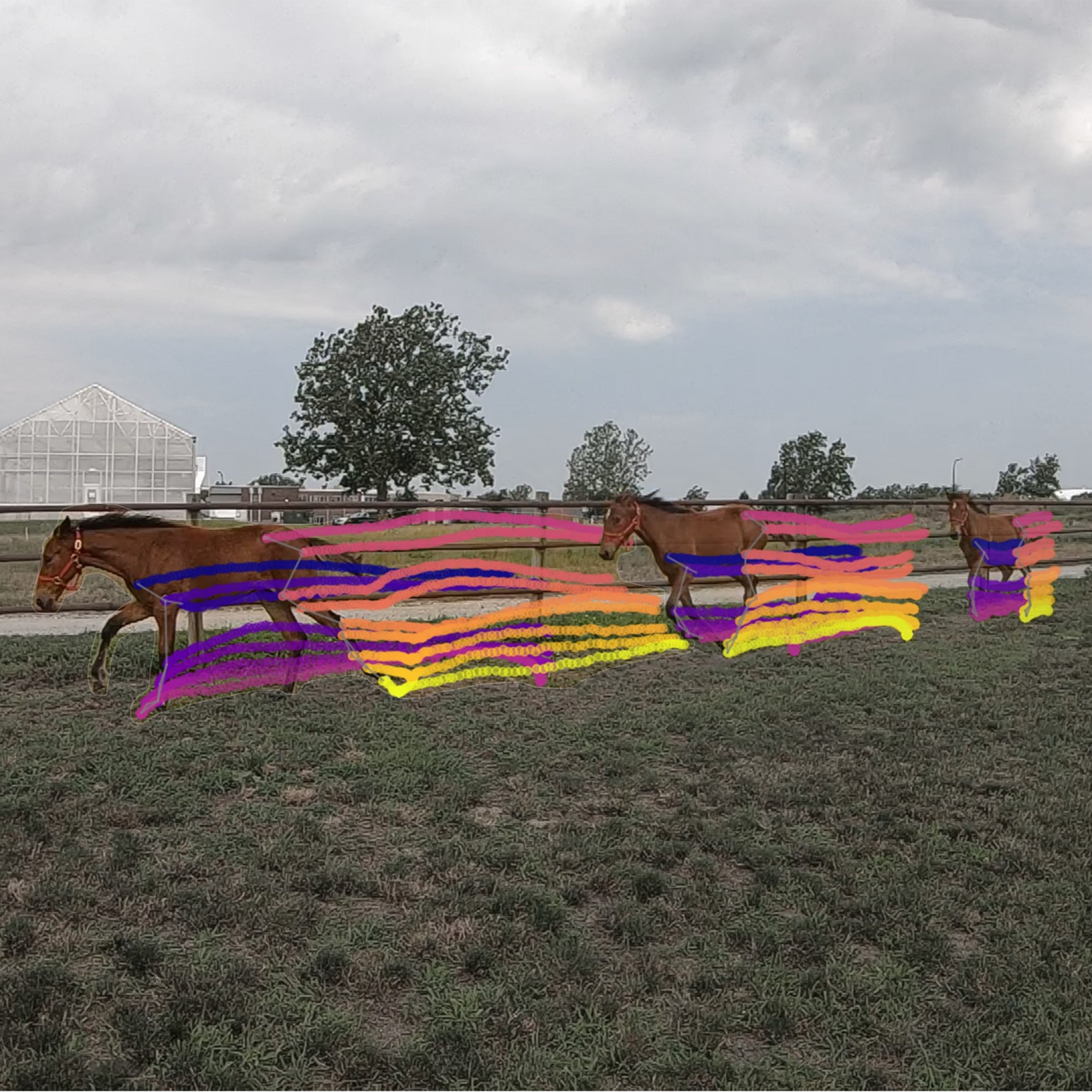 | Bone Properties During Growth and Fracture Prevention in Racehorses Most fatal musculoskeletal injuries in racing horses result from bone fracture. Specifically, these musculoskeletal injuries most often occur in the metacarpophalangeal (MCP) joint. The objective of this study is to characterize how equine bones in the lower limb adapt to changing loads during growth and exercise. We use longitudinal computed tomography (CT) scans, motion capture, force measurenemnts, and machine learning to accomplish this. Using the CT scans, we build subject-specific finite element models to measure bone strength in high-risk regions. Contributors: Melany Opolz, Sara Moshage, Kellie Halloran Collaborators: Professor Annette McCoy (UIUC, Veterinary Medicine), Professor John Polk (UIUC, Anthropology) Funding: UIUC VetMed Companion Animal Research Foundation, UIUC VetMed Hatch Grant, Morris Animal Foundation, Campus Research Board, Grayson Jockey Club |
||
 | Exercise and Bone Remodeling Exercise interventions may provide a feasible means for improving the strength of regions that are at high risk for fracture, however bone adaptation is sensitive to the mechanical loading from different exercises and postures. It remains unclear how exercise affects bone and whether it is at all beneficial to bone strength. We use ovine bone to investigate whether the mechanical properties of bone after a sixty-day exercise trial are significantly different from non-exercised controls. Specifically, we computed tomography (CT) scan bone samples, and afterwards we mechanically test the samples in compression. Contributors: Joshua Craggette, Sony Manandhar, Sara Moshage, Hyunggwi Song Collaborators: Professor John Polk (UIUC, Anthropology) Funding: National Science Foundation |
||
 | Effects of Fatigue and Stress Fracture Prevention in Athletes Bone serves a mechanical role to protect and support the musculoskeletal system over a lifetime of loading. However, repeated loading increases the risk of stress fracture, particularly among basketball players and military personnel. We study the tibia, one of the bones most prone to stress fractures, during basketball activities. Our objective is to develop preventive and management strategies to minimize fracture risk. We use motion capture, computed tomography (CT), and finite element (FE) modelling in this study. Contributors: Chenxi Yan Collaborators: Professor Stuart Warden (IUPUI, Physical Therapy) Funding: National Basketball Association, GE |
||