
Sara graduated and will be doing bioengineering consulting with Exponent in Philadelphia starting later this fall. Congratulations Dr. Moshage!! You will be missed!
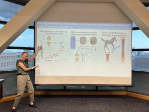
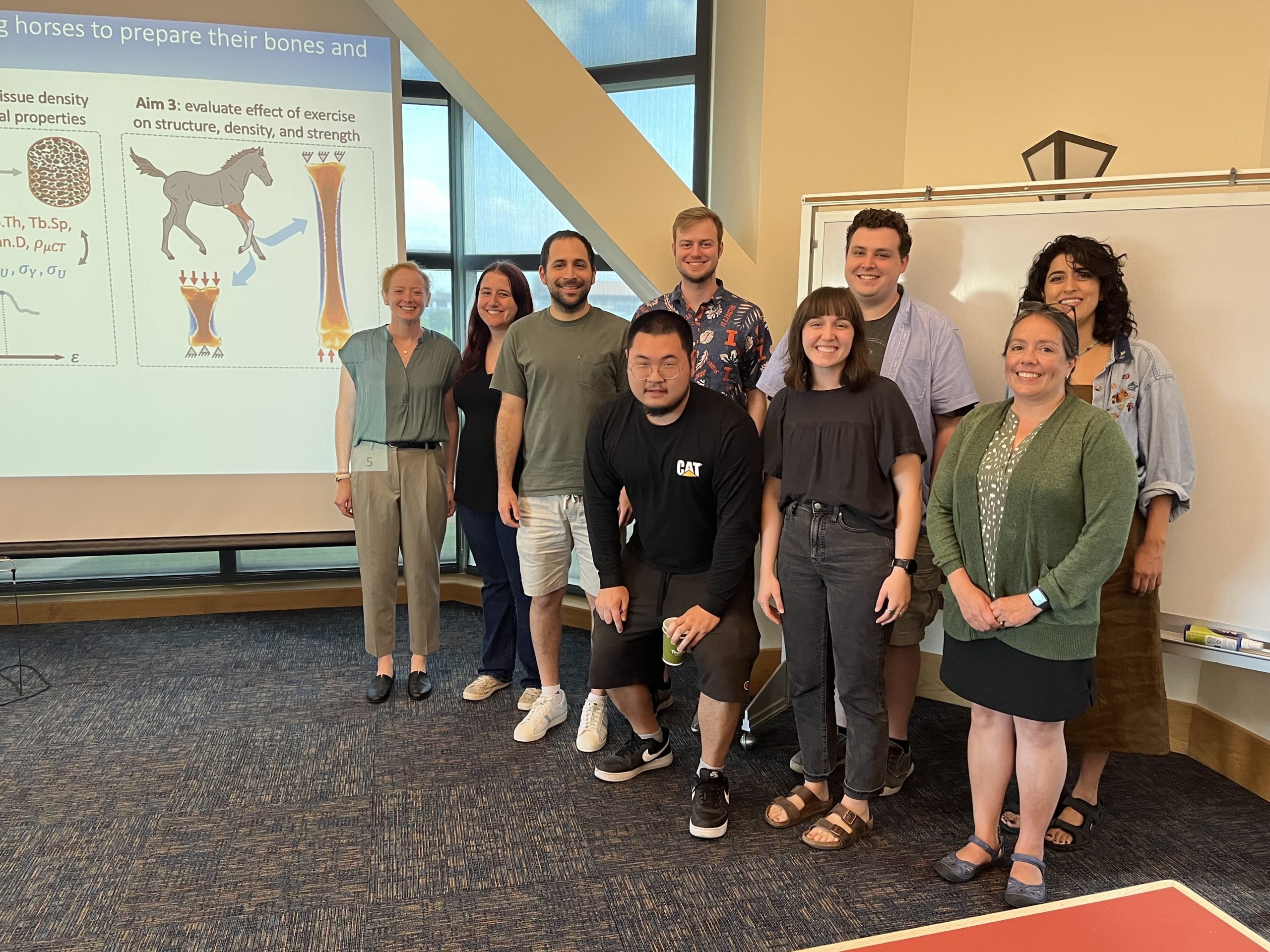
Summary of Sara’s Research
Fractures are the leading cause of racehorse fatalities, and more than 60% of fractures in the lower limbs occur in the third metacarpal (MC3) and proximal phalanx (P1) bones, and thus are the focus are this thesis. Exercise at a young age has been shown to have lifelong benefits for bone health, and may provide a route to strengthen bones in areas prone to fracture and prevent fractures in racehorses. Computational models can be used to non-invasively predict the mechanical loading environment of bone in vivo and therefore provide a means for evaluating the effect of different exercise modes pre-clinically rather than adopting a trial and error approach. Subject-specific finite element models can be developed from computed tomography (CT) data using a density-modulus relationship that relates CT-derived density to a modulus obtained from mechanical tests, but there is no density-modulus relationship for juvenile equine bone. In Aim 1, I found that the density-modulus relationships differed significantly by anatomical location (MC3 vs P1) and orientation (longitudinal vs trabecular), indicating each bone and orientation required a unique density-modulus relationship. In Aim 2, I developed relationships between mechanical properties, microstructure, and tissue density for MC3 and P1 trabecular bone that can be used for in vivo predictions of bone health, or to evaluate the effects of interventions (exercise or pharmacological) on trabecular microstructure of juvenile horses. In Aim 3, I evaluated the effects of an exercise intervention on MC3 and P1 structure, density, and strength using repeated CT scans and virtual compression testing in a group of juvenile horses. Overall, this thesis lays the foundation for using finite-element models to non-invasively assess exercise interventions in juvenile horses by (1) providing the necessary density-modulus relationships that accurately describe juvenile bone, (2) highlighting the influence of microstructure and tissue density on mechanical behavior and how those relationships change between neighboring anatomical locations, and (3) evaluating an exercise intervention using longitudinal CT scans and finite-element modeling.